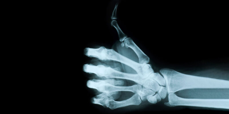
In the field of medical imaging, X-ray and DEXA scans are vital for diagnosing and planning treatments. Though both offer valuable information about the body, they have different uses and methods. Both healthcare providers and patients need to know the strengths and weaknesses of each. In this blog, we’ll explore the details of X-ray and DEXA scans, highlighting what they’re good at and where they fall short.
X-Ray Imaging: A Timeless Diagnostic Tool
X-ray imaging, discovered in 1895 by Wilhelm Conrad Roentgen, remains a widely used diagnostic tool in medicine. It uses radiation to create images of internal structures, especially bones and dense tissues. X-ray in Liverpool are great for finding fractures, joint problems, and issues in the skeletal system. They’re also crucial for diagnosing conditions such as pneumonia, tuberculosis, and lung diseases by showing the lungs and nearby tissues.
Pros of X-Ray:
-
Versatility:
X-rays are incredibly useful because they can take pictures of many body parts, which helps diagnose lots of different conditions, like dental problems and issues with the stomach and intestines. They’re also used in mammography to screen for breast cancer and in angiography to see blood vessels.
-
Speed:
X-ray imaging is fast, giving quick results that help diagnose and treat patients promptly. This speed is especially important in emergencies, where quick diagnosis is vital for managing patients.
-
Cost-effective and Widely Accessible
X-ray imaging offers a dual advantage of cost-effectiveness and wide availability. Being more economical compared to advanced imaging techniques, X-rays are accessible in numerous healthcare facilities. Their widespread presence, including in remote regions, ensures swift access to diagnostic services for individuals from all walks of life, regardless of geographical barriers or financial constraints.
Cons of X-Ray:
-
Limited soft tissue contrast:
X-rays are good at showing bones but not as good at showing soft tissues, which can make it harder to diagnose things like tumours or muscle injuries. To get a better look at soft tissues, doctors might use other imaging methods like MRI or CT scans.
-
Radiation exposure:
X-rays use radiation, which can be risky for health, especially with repeated or high doses. Even though modern X-ray methods use low doses to lower risks, healthcare providers need to be careful, especially with children and pregnant people.
-
Lack of specificity:
X-ray images might not show all the details, so additional tests or clinical evaluation may be needed for an accurate diagnosis, especially when abnormalities are subtle or structures overlap.
DEXA Scan: Precision in Bone Density Assessment
Unlike X-rays, DEXA scans focus on accurately measuring bone mineral density (BMD). Introduced in the 1980s, DEXA uses two different X-ray energy levels to measure BMD at certain places in the body, usually the spine, hip, or forearm. This method is crucial for diagnosing osteoporosis, a condition marked by lower bone density and higher risk of fractures, by evaluating a person’s bone health and risk of fractures.
Pros of DEXA Scan:
-
High precision:
DEXA scans are incredibly precise in measuring bone mineral density, which helps diagnose and track osteoporosis and similar conditions accurately. By measuring BMD carefully in important bone areas, a DEXA scan can spot bone loss early and help decide on the right treatments to avoid fractures.
-
Minimal radiation exposure:
DEXA scans expose patients to very little radiation, reducing worries about health issues, especially for vulnerable groups. This low radiation dose in DEXA scans lowers the risks associated with radiation while still giving important details about bone health.
-
Site-specific analysis:
DEXA scan analyses bone density in specific areas, helping doctors identify weak spots and plan treatments accordingly. By looking at BMD in places like the spine or hip, DEXA scans give important information about fracture risk and help decide on the best ways to keep bones healthy.
Cons of DEXA Scan:
-
Limited soft tissue evaluation:
Although DEXA scans are good at measuring bone density, they don’t give much detail about soft tissues or other structures, which could mean missing other problems. To fully check for musculoskeletal issues and soft tissue abnormalities, additional imaging methods like MRI or ultrasound are needed.
-
Cost and accessibility:
Not all healthcare places have DEXA scanners and trained staff, which can make it hard for some patients in certain areas to get access. The cost of DEXA scans might also stop people from getting them, especially in areas with fewer resources.
-
Interpretation challenges:
Understanding DEXA scan results needs special training because things like age, sex, and ethnicity can affect bone density readings, so doctors need to be careful when interpreting them. They have to think about different things specific to each patient when looking at DEXA results and deciding what to do about osteoporosis.
Conclusion
X-ray and DEXA scans are different but work well together, each with its strengths and weaknesses. X-rays are great for seeing bones and finding many problems, while DEXA scans are best for measuring bone density, especially in osteoporosis diagnosis. Knowing what each method is good at and where it might not be as helpful is crucial for doctors to decide on the best imaging and care for patients. As technology gets better, using both of these methods together can improve diagnosis and help patients even more in today’s changing medical world.
Remember, prioritising your health shouldn’t come at the cost of financial burden. CareScan offers exceptional radiology services at fair prices, ensuring you get the care you need without financial strain. Contact them today for affordable diagnostic tests and the best treatments.

Comments